Brain scans are powerful tools that allow doctors and researchers to see inside the brain without the need for surgery. These scans help diagnose medical conditions, understand brain function, and guide treatment plans. There are three main types of brain scans: MRI, CT, and PET scans. Each type uses different technology and provides unique information about the brain. Let’s explore these three types of brain scans in detail.
Magnetic Resonance Imaging (MRI) Scans
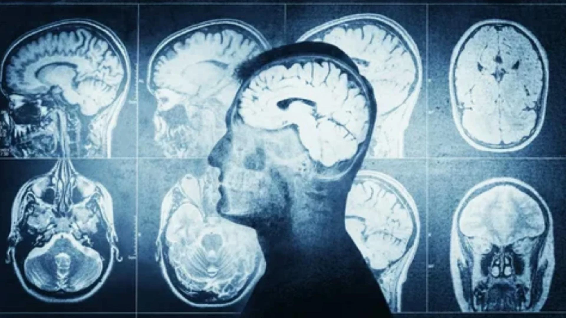
Magnetic Resonance Imaging, commonly known as MRI, is one of the most detailed and widely used brain imaging techniques. An MRI scan uses strong magnetic fields and radio waves to create detailed images of the brain’s structure. Unlike other imaging methods, MRI does not use radiation, making it a safer option for many patients.
The process of an MRI scan involves lying inside a large, tube-shaped machine. The machine generates a magnetic field that aligns the protons in your body’s water molecules. Radio waves are then sent through the body, causing the protons to produce signals. These signals are detected by the machine and converted into highly detailed images of the brain.
One of the key advantages of an MRI scan is its ability to produce high-resolution images of soft tissues, such as the brain. This makes it particularly useful for detecting abnormalities like tumors, bleeding, or inflammation. MRI scans are also commonly used to study the brain’s anatomy and diagnose conditions such as multiple sclerosis, strokes, and brain injuries.
There are different types of MRI scans, including functional MRI (fMRI), which measures brain activity by detecting changes in blood flow. This type of scan is often used in research to understand how different parts of the brain work during specific tasks or in response to stimuli. Overall, MRI scans are a versatile and powerful tool for examining the brain.
Computed Tomography (CT) Scans
Computed Tomography, or CT scans, are another common type of brain imaging. CT scans use X-rays to create cross-sectional images of the brain. Unlike a standard X-ray, which produces a single image, a CT scan takes multiple images from different angles. These images are then combined by a computer to create a detailed, three-dimensional view of the brain.
During a CT scan, the patient lies on a table that slides into a doughnut-shaped machine. The machine rotates around the head, taking X-ray images from various angles. The entire process is quick, often taking only a few minutes, making it a convenient option for emergencies.
CT scans are particularly useful for detecting structural abnormalities in the brain, such as skull fractures, bleeding, or swelling. They are often used in emergency situations, such as after a head injury, because they provide fast and accurate results. CT scans are also helpful in diagnosing conditions like strokes, brain tumors, and infections.
One of the main advantages of CT scans is their ability to provide clear images of both bone and soft tissue. However, they do involve exposure to a small amount of radiation, which is something to consider, especially for patients who may need multiple scans over time. Despite this, CT scans remain a valuable tool for diagnosing and monitoring brain conditions.
Positron Emission Tomography (PET) Scans
Positron Emission Tomography, or PET scans, are a type of brain imaging that focuses on brain function rather than structure. PET scans use a radioactive tracer, which is injected into the bloodstream, to measure activity in different parts of the brain. The tracer collects in areas of the brain that are more active, allowing doctors to see how the brain is working.
During a PET scan, the patient is injected with the tracer and then lies on a table that slides into a scanning machine. The machine detects the radiation emitted by the tracer and creates images that show brain activity. These images can reveal how well the brain is functioning and identify areas of abnormal activity.
PET scans are particularly useful for diagnosing and monitoring conditions that affect brain function, such as Alzheimer’s disease, epilepsy, and Parkinson’s disease. They can also be used to evaluate the effectiveness of treatments, such as chemotherapy for brain tumors. In addition, PET scans are often used in research to study brain disorders and understand how different parts of the brain interact.
One of the unique aspects of PET scans is their ability to provide information about brain metabolism and chemistry. This makes them a valuable tool for understanding complex brain conditions that may not be visible on structural scans like MRI or CT. However, because PET scans involve radiation, they are typically used when other imaging methods cannot provide the necessary information.
Comparing the Three Types of Brain Scans
Each type of brain scan has its own strengths and limitations, and the choice of scan depends on the specific needs of the patient. MRI scans are ideal for detailed images of soft tissues and are often used for diagnosing structural abnormalities. CT scans are faster and better suited for emergencies, providing clear images of both bone and soft tissue. PET scans, on the other hand, focus on brain function and are useful for diagnosing and monitoring conditions that affect brain activity.
In some cases, doctors may use a combination of scans to get a complete picture of the brain. For example, a patient with a suspected brain tumor might undergo a CT scan to quickly assess the situation, followed by an MRI for more detailed images, and a PET scan to evaluate the tumor’s activity. By combining the strengths of different imaging techniques, doctors can make more accurate diagnoses and develop effective treatment plans.
Conclusion
Brain scans are essential tools for understanding and diagnosing brain conditions. The three main types of brain scans—MRI, CT, and PET—each offer unique insights into the brain’s structure and function. MRI scans provide detailed images of soft tissues, CT scans are fast and effective for emergencies, and PET scans focus on brain activity and metabolism. By using these imaging techniques, doctors can better diagnose and treat a wide range of brain-related conditions, improving patient outcomes and advancing our understanding of the brain. Whether you’re a patient or a researcher, understanding these three types of brain scans can help you appreciate the incredible technology that allows us to explore the most complex organ in the human body.